Optical Microscope vs Scanning Electron Microscope
Optical Microscope have been in existence since the 13th century. Earliest compound microscopes are believed to have appeared in 1620 whereas even the first electron microscope (TEM) was invented in 1931. If optical microscopes (OM) had been in existence since such a long time, why was then there a need for an electron microscope (EM)?
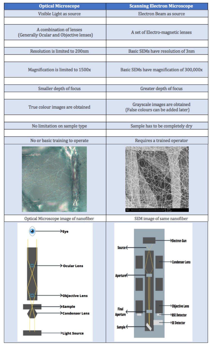
OM use visible light as a source for imaging and inspection and a system of lenses to generate magnified images of small objects.
OM use visible light as a source for imaging and inspection and a system of lenses to generate magnified images of small objects.
SEM on the other hand uses electrons as a source and a combination of electro-magnetic lenses to generate magnified images of the sample.
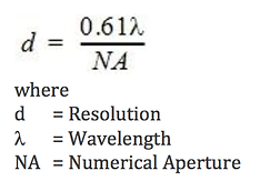
In the field of microscopy, resolution is the most important parameter to consider. Resolution here is defined as the shortest distance between two points on a specimen that can be clearly distinguished by the observer as separate entities. The wavelength of the source directly influences the resolution of a microscope. In optical microscopy, the resolution is limited by low wavelength of visible light (400 – 700nm). Because of this, these microscopes can offer a 1500x magnification only and a minimum resolution of 200 nm.
The electron microscope was developed when the wavelength became the limiting factor in light microscopes. The beam used in SEMs contains electrons with energies thousand times greater than that of visible light. The wavelength in SEMs are much shorter and hence they have much better resolving power. A standard tungsten source SEM can have resolution of order of 3.0nm with magnification ranging around 300,000x.