Notification

CONFOCAL RAMAN MICROSCOPE

Raman microscopy couples a Raman spectrometer to a standard optical microscope, allowing high magnification visualization of a sample and Raman analysis with a tiny laser spot. Raman microscopy is easy: place the sample under the microscope, focus, and make a measurement.
Just adding a microscope to a Raman spectrometer does not give a controlled sampling volume - for this a spatial filter is required. Confocal Raman microscopy refers to the ability to spatially filter the analysis volume of the sample, in the XY (lateral) and Z (depth) axes.
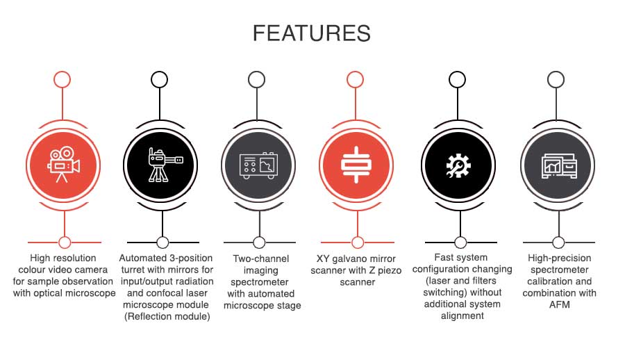
RAMOS E/M Series Raman spectrometers are designed on the basis of research-grade optical microscopes allowing the realization of the following light microscopy methods:
- Raman measurements
- Transmitted light
- Reflected light (bright field and dark field illumination)
- Confocal microscopy
- Fluorescence measurements
- Polarisation contrast and phase contrast imaging
- Differential interference contrast